Is It Possible For The Body To Repair A Small Leak In Amniotic Fluid
Abstract
Preterm premature rupture of membrane (pPROM) is associated with xxx–40% of preterm births. Infection is considered a leading cause of pPROM due to increased levels of proinflammatory cytokines in amniotic fluid. Just 30%, however, are positive for microbial organisms by amniotic fluid culture. Interestingly, in some pregnancies complicated by preterm premature rupture of membranes (pPROM), membranes heal spontaneously and pregnancy continues until term. Here, we investigated mechanisms of amnion healing. Using a preclinical mouse model, nosotros found that pocket-sized ruptures of the fetal membrane airtight within 72 h whereas healing of large ruptures was only forty%. Small rupture induced transient upregulation of cytokines whereas big ruptures elicited sustained upregulation of proinflammatory cytokines in the fetal membranes. Fetal macrophages from amniotic fluid were recruited to the wounded amnion where macrophage adhesion molecules were highly expressed. Recruited macrophages released limited and well-localized amounts of IL-1β and TNF which facilitated epithelial-mesenchymal transition (EMT) and epithelial cell migration. Arg1 + macrophages dominated within 24 h. Migration and healing of the amnion mesenchymal compartment, however, remained compromised. These findings provide novel insights regarding unique healing mechanisms of amnion.
Introduction
Preterm labor is the leading crusade of perinatal morbidity and mortality1. Preterm premature rupture of membrane (pPROM) is defined equally the rupture of membrane occurring before 37 weeks of gestation, which is associated with xxx–40% of preterm deliveries and occurs in approximately 1–three% of all pregnanciesii. Amniotic fluid cultures signal that 30% of pPROM are positive for microbial organismsiii. Infection-related pPROM requires firsthand intervention (delivery) for fright of infection to fetus such as fetal inflammatory syndrome, which is a take a chance of severe neonatal morbidity with respiratory distress syndrome, neonatal sepsis, pneumonia, chronic lung disease, necrotizing enterocolitis, intraventricular hemorrhage, and cognitive palsy4, as well as maternal complication such as sepsis. On the other paw, the majority of pPROM cases are unrelated to infection but may be associated with smoking, depression trunk mass-index, maternal stress, and intrauterine bleeding. In add-on, iatrogenic pPROM is caused past amniocentesis or fetoscopy, and accidental rupture of membrane during surgery such as cervical cerclage. It is mostly idea that rupture of membrane is irreversible upshot because most women with pPROM begin labor spontaneously within several days. A small proportion, yet, remain undelivered2 with spontaneous sealing of the membranesv. The mechanisms that promote healing of the fetal membranes are unknown.
Macrophages play an of import part in wound healing6,seven. Macrophages phagocytose pathologic organisms and matrix debris, removing necrotic tissue. Macrophage also releases diverse growth factors and cytokines at the wound site, reconstituting the wound site past forming new blood vessels and regulating fibroblast recruitment. Although gradations occur in macrophage classifications, in general, macrophages differentiate into 2 phenotypes: Classical interferon-γ (IFN-γ)-activated macrophages (M1 macrophages) past T helper 1 (Th1)-type responses play a role of cellular immunity to infections whereas culling activated macrophage (M2 macrophages) activated by Th2-type cytokines IL-four and IL-13 are important in allergic reactions, parasitic infections, fetal tolerance, and tissue repaireight,9. M2 macrophages exhibit stiff anti-inflammatory activeness and serve important roles in wound healing. Furthermore, M2-like macrophages antagonize proinflammatory M1-macrophage responses10.
Fetal membranes are comprised of amnion, chorion, and decidua. Amnion is the primary load-bearing structure of the fetal membranes1 and is composed of two jail cell types: superficial epithelial cells and underlying mesenchymal cells. Interstitial collagens (types I, 3, and V) are produced by amnion mesenchymal cells to maintain the mechanical integrity of the amnion. Protected inside these durable fetal membranes, amniotic fluid serves a defensive role every bit an innate allowed systemxi, and a reservoir for fetal macrophages12. Maternal macrophages are enriched in maternal-derived deciduaxiii.
Here we investigated the mechanism of wound healing using sterile rupture of the fetal membranes in preclinical mouse models (small and large ruptures). Whereas small-scale ruptures of the amnion closed past 72 h, > fifty% of large ruptures remained open up. Macrophages from amniotic fluid were recruited to the wounded amnion where macrophage adhesion molecules were highly expressed. Initially, recruited macrophages released limited and well-localized amounts of IL-1β and TNF. These proinflammatory cytokines facilitated epithelial-mesenchymal transition (EMT) in human amnion epithelial cells, and EMT was also observed in the healing amnion of mice. Arg1-positive macrophages were dominant within 24 h. Using this mouse fetal membrane rupture model, we conclude that fetal membranes heal by unique mechanisms with compromise of amnion mesenchymal cell migration in big ruptures.
Results
Preclinical mouse model of sterile ruptured membrane
A mouse model of sterile rupture of the fetal membranes was generated using puncture of the amniotic membrane with 26 or xx G needles (outer diameter 0.47 or 0.91 mm) at xv embryonic day (ED). By puncture, amniotic fluid leaked through myometrium (Fig. 1A) and in the vagina through the cervix (Fig. 1B). After removal of myometrium, ruptured edge of choriodecidua was readily visible (Fig. 1C,D), and long centrality of rupture was measured. Edge of amnion was commonly stained with black ink (Fig. 1C) thereby distinguished from choriodecidua. Long axis of amnion rupture was likewise measured (Fig. 1D). For cases in which rupture of amnion was unclear, choriodecidua was gently slid with cotton wool swab to clarify edge of ruptured amnion. If amnion was healed completely, stain of amnion looked similar a dot (Fig. 1E). 2 h after rupture, perforations were macroscopically visible (Fig. 2A, 2 h). At 24 h, almost half of injuries were however visible, but perforation bore decreased significantly. Rupture was closed later 48 and 72 h (Fig. 2A). Histologic analysis of the rupture site in transverse cross-department revealed that membrane structure was interrupted 2 h after rupture (Fig. 2B, 2 h). After 24 h, amnion thickness increased at the edge of injury (Fig. 2B, 24 h). In our model, 26 K-induced perforations of amnion were almost closed past a monolayer of epithelial cells roofing stratified mesenchymal cells within 48–72 h (Fig. 2B, 24–72 h).
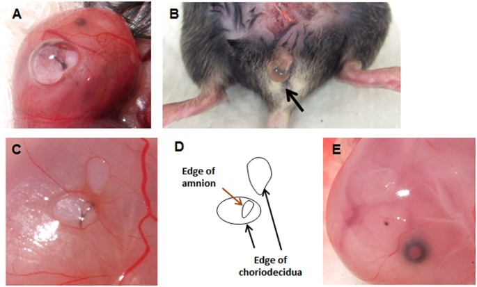
Mouse model of sterile ruptured membranes with 26 estimate (ø 0.47 mm) needle. Leakage of amniotic fluid after puncture through fetal membrane (A) and into vagina through neck (B). (C) Ruptured fetal membrane 24 h after rupture with 20 ga needle. Myometrium has been removed to visualize membrane defects. Annotation that edge of ruptured amnion was stained with black ink. (D) Schematic of panel C illustrating choriodecidual and amnion defects. (E) Completely healed amnion and choriodecidua 72 h after rupture with 26 ga needle.
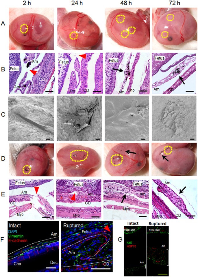
Preclinical mouse model of sterile ruptured membranes. (A–F) Time class of amnion healing. (A) Macroscopic images of ruptured fetal membranes with 26 G needle. Ruptured site was stained with Bharat ink generating blackness staining at the center of circles outlined in yellowish. (B) Hematoxylin-Eosin (H&E) staining of ruptured fetal membranes by 26 Thousand needle (x 400). Whole gestational sac including myometrium, fetal membranes, and fetus was stock-still and syectioned at the site of injury. Am, amnion; Cho, chorion; December, decidua; CD, choriodecidua; Myo, myometrium. Ruby arrowhead indicates the defect site of amnion and black arrow indicates healed site. Annotation that ruptured site was stained with blackness India Ink. Bars, 50 µm. (C) Images of scanning electron microscopy (SEM). Afterwards removal of fetus and placenta, images of fetal membranes were captured from inside the gestational sac. Hence, the surface shown here is the epithelial layer of amnion. Bars, 20 µm. (D–East) Sterile ruptured membrane with 20 gauge (ø 0.91 mm) needle. (D) Macroscopic images of ruptured fetal membranes at indicated fourth dimension subsequently 20 G rupture. Yellow dashed lines encircle ruptured sites stained with black India Ink. Yellowish dashed lines encircle open up puncture wounds (not healed) whereas pointer indicates closed healing sites. (E) H&East staining of fetal membranes after xx Grand rupture at indicated time. Bars, 50 µm. (F) Immunofluorescence staining for vimentin (light-green), E-cadherin (blood-red), and DAPI (blue) of fetal membranes from intact site on embryonic d15 (Intact) or border of healing amnion 24 h subsequently 20 Yard rupture (Ruptured). Amnion is circled by white dash lines. Bars, 50 µm. (Yard) Immunolocalization of HSP70 (red) and Ki67 (green) in intact (not-ruptured) and healing ruptured membranes 24 h after 20 G puncture. Note proximity of thickened amnion to fetal skin compared with intact nonruptured membrane. Bar, 100 µm.
Scanning electron microscopy (SEM) was used to clarify the ultrastructure of healing fetal membranes. Images were taken from within the gestational sac, and so that images reflect the inner surface of amnion lining the gestational sac. Interestingly, collapsed perforations with surgically-precise edges were readily observed 2 h after rupture (Fig. 2C, two h). At 24 h, amnion prison cell migration and matrix deposition was noted in the rupture site, generating a plug- or flap-like structure (Fig. 2C, 24 h). At 48 and 72 h, the rupture site was closed with thickening and bulging, presumably reflecting migration of mesenchymal cells and matrix deposition as seen by light microscopy (Fig. 2C, 48 h and 72 h). In addition, the ruptured site appeared to be covered by amnion epithelial cells (Fig. 2C, 48 h and 72 h, higher resolution images are shown in Fig. S1A,B).
Next, we compared rapid complete closure of 26 G punctures with larger 20 Thousand ruptures (outer bore, 0.91 mm). In contrast to near 100% healing of modest ruptures, almost half of 20 K rupture sites remained open even after 72 h (Fig. 2d). Histologic analysis of the rupture sites revealed accumulation of amnion mesenchymal cells (Fig. 2E, 48 h and 72 h). SEM images of the large rupture model were non possible due to adherence of the ruptured membrane to the fetus from severe loss of amniotic fluid. Thus, after fixation, the membrane was fragile and destroyed during removal of the fetus from the gestational sac. To report the dynamics of healing of 20 Thou rupture sites, immunofluorescence studies were conducted with markers of epithelium and mesenchyme. In intact fetal membranes, the epithelial cell marker E-cadherin was predominantly localized at cell-cell junctions of amnion epithelial cells and chorion (Fig. 2F). Staining was highly localized and organized at regular intervals between flattened epithelial cells. Vimentin-positive amnion mesenchymal cells were localized to 1-2 cell layers beneath the epithelial lining (Fig. 2F, intact), and fibroblasts in the chorion were also vimentin-positive (Fig. 2F, intact). In the injured epithelium, Due east-cadherin staining was localized diffusely throughout the cell membrane within 24 h (Fig. 2F, ruptured). Vimentin-positive amnion mesenchymal cells were prominent at the rupture site resulting in a thickened edge of healing amnion (Fig. 2F, ruptured).
To determine if this thickened border of healing amnion was due to proliferation, Ki67 immunostaining was colocalized with HSP70 to ensure right localization of the injury site (Fig. 2G). In not-ruptured membranes, HSP70 was punctate and sparsely distributed. In the ruptured membrane after 24 h, HSP70 was more intense and widely distributed throughout the thickened amnion which is closer to the fetal skin due to loss of amniotic fluid (Fig. 2F). Interestingly, Ki67 was non increased in the injured amnion relative to intact membranes and less than the robust Ki67 staining observed in proliferating fetal skin (Fig. 2G).
The average diameter of small ruptures was 0.12 mm at 24 h and 0.03 mm at 72 h whereas in large ruptures, diameters of 1.25 mm at 24 h (increased significantly compared with pocket-sized rupture at 24 h) decreased to 0.82 mm at 72 h (P < 0.01 compared with modest rupture at 72 h) (Fig. 3A). In the small rupture model, boilerplate complete closure of amnion was 83% at 24 h and 98% at 72 h (Fig. 3B). In the large rupture model, however, closure rates were only 7% at 24 h and 48% at 72 h (Fig. 3B). In choriodecidua, the average diameter of modest rupture was 0.31 mm at 24 h and 0.24 mm at 72 h (statistically not significant), whereas in large rupture, healing was remarkably delayed: 1.38 mm at 24 h (P < 0.01 compared with small rupture) and 1.29 mm at 72 h (P < 0.01 compared with small rupture, Fig. S2A). Closure of choriodecidua was impaired significantly relative to amnion with 61% and 78% at 24 and 72 h, respectively in small rupture model (Fig. S2B). In the large rupture model, the choriodecidua did not heal at 24 h (no closures) and just xvi% at 72 h (Fig. S2B).
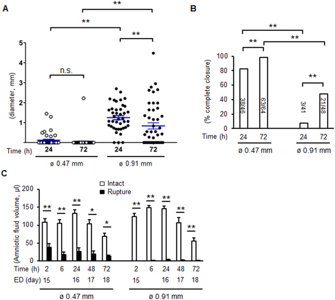
Healing rate and perforation diameters of amnion after membrane puncture. (A) Diameter of ruptured membrane sites in amnion with 26 G (ø 0.47 mm) or twenty G needle (ø 0.91 mm) at 24 and 72 h of puncture. Each symbol represents one rupture. Blue bar indicates mean and SEM. **P < 0.01, ANOVA. (B) Percent consummate closure of ruptured amnion at 24 and 72 h. Number of completely closed ruptures/total ruptures is shown as the bar. *P < 0.05, *P < 0.01, χii. northward = 41–64 punctures from 12–17 fetal membranes of 3–seven significant mice in each group. (C) Amniotic fluid volume afterward rupture with a 26 G (ø 0.47 mm) or twenty M needle (ø 0.91 mm). ED, embryonic solar day. Values were compared to the volume of intact gestational sac at each time point. Error confined represent SEM. due north = half-dozen–20 gestational sacs from three–vii pregnant mice in each grouping. *P < 0.05, **P < 0.01.
At the time of puncture, pregnant amounts of amniotic fluid leaked from the gestational sac with increased losses in the large rupture model (Fig. 3C). Interestingly, although puncture wounds closed in the small-scale rupture grouping, amniotic fluid volume did not recover at 48 and 72 h. Despite significant decreases in amniotic fluid, intrauterine fetal survival rates were non decreased after small rupture (>86%) just compromised somewhat by big rupture at 72 h (from 100 to 82%, P < 0.05, χtwo). This increased fetal loss appeared to be prolapse of the umbilical cord through the ruptured site. The majority of fetuses appeared healthy with normal fetal growth despite oligohydramnios (fetal weight: 1.06 g in intact, one.00 one thousand in ø 0.47 mm, and one.01 chiliad in ø 0.91 mm, n = 4–six in each group). Farther, markers of lung alveolar epithelium differentiation were like in fetal lung from intact and ruptured membranes on d18.5 (Supplementary Tabular array two).
Unique factor expression profiles of wound healing in the injured amnion
Classically, developed wound healing is comprised of iv steps: (1) hemostasis, (2) inflammation, (iii) proliferation and (4) remodeling6,7,14. Injured vessels play a major office in this process from the early process of clot formation, immune jail cell recruitment, and release of growth factors from neoangiogenesis. The unique nature of the avascular amnion comprised of fetal cells suggests that basic wound-healing mechanisms must differ in fetal membranes. To investigate these mechanisms, factor expression profiles related to wound healing were quantified in fetal membranes with or without injury (Fig. 4). Using fetal membranes in vivo (samples that include amnion, chorion, decidua, and all cells attached to membrane), mRNA levels of proinflammatory cytokines, interleukin-1β (Il1b), tumor necrosis factor (Tnf), and Il6 increased within 2 h later small-scale puncture, returning to normal levels by 24 h. In dissimilarity, after large rupture, relatively loftier levels of Il1b, Tnf, and Il6 mRNA persisted until 72 h. Interestingly, the anti-inflammatory cytokine, Il10, was also remarkably upregulated in ruptured membranes, especially after large rupture (sixty-fold compared with eight-fold for Tnf, Fig. four).
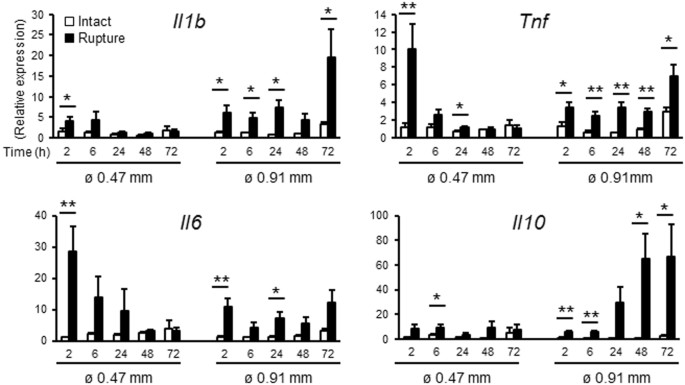
Rupture of fetal membranes alters inflammatory cistron expression in vivo. Expression of Il1b, Tnf, Il6, and Il10 in intact or ruptured (ø 0.47 mm: 26 G needle with viii punctures/sac, and ø 0.91 mm: xx G needle with 4 punctures/sac) fetal membranes. Relative mRNA expression of each gene normalized to that of βii microglobulin was compared to levels of intact membrane at two h. For statistical analysis, gene expression in ruptured membrane was compared with that of intact membrane at each fourth dimension point. Error bars represent SEM. n = 5–six fetal membranes from 5-half dozen significant mice in each group. *P < 0.05, **P < 0.01.
Growth factors play an important function in wound healing of many adult tissues. In the healing of fetal membranes, however, mRNA levels of these growth factors (epidermal growth factor, Egf; basic fibroblast growth factor, Bfgf) or Fgf2; transforming growth factors, Tgfb1 and 3; vascular endothelial growth factor, Vegf; and insulin-like growth factors, Igf1 and two) were not induced by sterile rupture (with the exception of a slight increase in Tgfb2 after big rupture) (Fig. S3). Since these growth factors are largely involved in angiogenesis, it is reasonable that they were not regulated in the avascular amnion. Even TGF-β2 did not stimulate migration of either epithelial or mesenchymal cells of amnion (information non shown). Overall, the results illustrate that amnion does not heal by the aforementioned mechanisms of other vascularized tissues.
Collagen matrix is remodeled in the later stage of wound healing. Hence, collagen germination at the healing site was compared using trichrome staining at 72 h and qPCR of collagen mRNA later 26 or twenty Thousand rupture. Later 26 Chiliad rupture, collagen fibers were thin forming a loose supportive network of the healed amnion (Fig. 5A) and mRNA levels of types 1, 3 and five collagen were non increased (Fig. 5C). In contrast, collagen deposition was dense with big deposits in the thickened healing sight afterwards 20 G rupture (Fig. 5B). Further, type ane and 3 collagen mRNA increased at 72 h (Fig. 5C). MMP9 mRNA was besides upregulated from 24 h in the large rupture (Fig. 5C). Collectively, agile collagen synthesis and matrix remodeling occurred in the healing membrane after large rupture. The lack of increased Ki67 at the wound site, together with vimentin-positive cells inside the thickened edge, suggests that healing of the wound is due to migration, EMT, and matrix deposition.
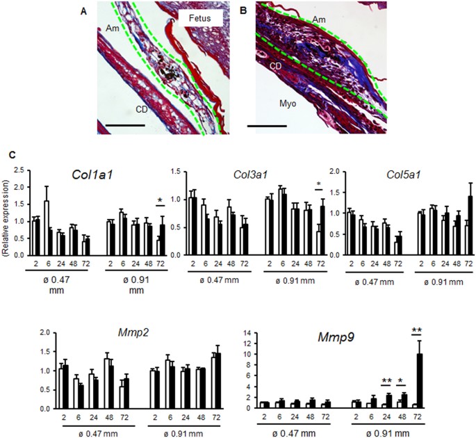
Collagen degradation in fetal membranes after pocket-sized or large rupture. Trichrome staining of ruptured fetal membrane 72 h later on puncture with 26 G (A) or 20 One thousand (B) needle. Amnion is indicated by greenish dashed lines. Bar, fifty µm. (C) Expression of Col1a1, Col3a1, Col5a1, Mmp2, and Mmp9 in intact or ruptured (ø 0.47 mm: 26 G needle with 8 punctures/sac, and ø 0.91 mm: 20 Yard needle with 4 punctures/sac) fetal membranes. Relative mRNA expression of each gene normalized to that of βii microglobulin was compared to levels of intact membrane at ii h. For statistical analysis, gene expression in ruptured membrane was compared with that of intact membrane at each time indicate. Error confined represent SEM. n = 5–6 fetal membranes from 5–6 pregnant mice in each group. *P < 0.05, **P < 0.01.
Recruitment of amniotic fluid macrophages in ruptured amnion
Innate amnesty assists wound healing, and macrophages play a central roleten. To investigate the interest of macrophages in healing of the ruptured fetal membrane, immunostaining of macrophages was conducted at the healing site of both small (Fig. S4B,I) and big ruptured amnion 24 h after injury (Figs 6B and S4I). Two macrophage markers (F4/fourscore and CD68) were utilized indicating similar results regarding macrophage number and location. Macrophages were almost absent in intact amnion before rupture (Figs 6A, S4A,I). In dissimilarity, macrophages localized to the amnion 24 h after injury (Figs 6B and S4B) with rare macrophages in chorion and decidua (Fig. 6C). In dissimilarity with other tissues in which neutrophils are recruited in the early stage of wound healing, neutrophils were rare in the healing amnion (Fig. S4C,D).
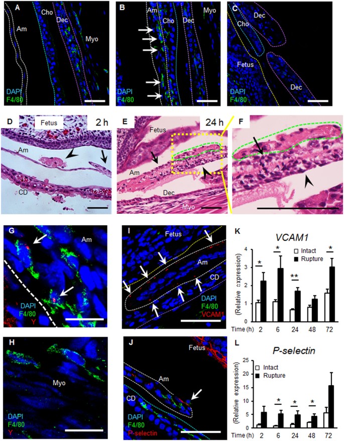
Macrophages and adhesion molecules at rupture site. (A–C) Immunostaining for F4/80 (light-green) and DAPI (blue) of membrane 24 h after rupture by 20 G needle. Dashed lines betoken amnion (Am, white), surface of fetal skin (xanthous), chorion (Cho, light blue), and decidua (December, imperial). Myo, myometrium. (A) Intact fetal membrane. (B) Macrophages inside healing amnion (arrows). (C) Ruptured edge of chorio-decidua. Bars, 50 µm. (D–F) H&E staining of ruptured membranes. (D) An amniotic fluid macrophage (arrow caput) attaches to ruptured amnion at 2 h (pointer). (Due east and magnified view in F) Amniotic fluid macrophages (green) attach to amnion surface at 24 h, and mesenchymal cells drift (arrow caput). Note mesenchymal-similar shape alter of epithelial cells in F. Bars, 50 µm. (G and H) Fluorescent in situ hybridization (FISH) of Y chromosome (carmine) and immunostaining for F4/fourscore (dark-green) at healing amnion (Grand) and intact myometrium (H) at 48 h. Bars, 10 µm. (I–L) Macrophage adhesion molecules at the ruptured site. Immunostaining for F4/80 (green), VCAM1 (I) or P-selectin (J, red) and DAPI (blue) at ruptured amnion at 24 h after rupture by 20 M needle. Confined, l µm. (K and 50) Gene expression of VCAM1 (Grand) or P-selectin (L) of intact or ruptured fetal membrane with xx Thousand needle at indicated fourth dimension. Error bars correspond SEM. n = 5–6 fetal membranes from 5–6 pregnant mice in each grouping. *P < 0.05, **P < 0.01.
Next, we sought to determine the origin of macrophages recruited to the site of injury. Macrophages fastened to the ruptured amnion as early as 2 h (Fig. 6D), and firmly attached to the ruptured site within 24 h (Figs 6E–F and 7C), with some incorporated within the amnion (Figs 6B,7 and S4). Remarkably, macrophage attachment was limited to the fetal side (Fig. 6D–F). In fetal membranes derived from male person fetus, macrophages incorporated in the healing amnion were Y-chromosome positive using fluorescent in situ hybridization (FISH, Fig. 6G), confirming that these macrophages were of fetal origin. In contrast, macrophages in myometrium were Y-chromosome negative (maternal origin, Fig. 6H).
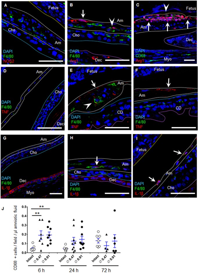
Localized cytokine secretion from M-2 macrophages. (A–C) Immunofluorescence staining of fetal membrane for F4/80 (green) or CD68 (light-green), NO synthase-2 (NOS2, red in A), arginase-one (Arg1, ruddy in B) and DAPI (blue) at 24 h of rupture by a xx 1000 needle (ø 0.91 mm). Dotted lines indicate amnion (white), surface of fetal skin (yellow), chorion (light blue), and decidua (regal). Note that Arg1 was expressed both in macrophages (B and C, arrowhead) and in amnion epithelial cells (B and C, arrow). Effigy 7C is same location equally Fig. 6E and F. Confined, fifty µm. (D–I) Immunofluorescence staining of fetal membrane for F4/80 (dark-green), TNF (crimson in D–F), IL-1β (red in G–I) and DAPI (blue) by a 20 G needle (ø 0.91 mm). (Eastward and F) TNF at healing site of amnion in macrophage (arrow head) and amnion epithelial cells (arrow) at 6 h after rupture. (D) Intact fetal membrane at 6 h. (H and I) IL-1β at healing amnion in macrophage (pointer) at 24 h after rupture. (G) Intact fetal membrane at 24 h. Confined, fifty µm. (J) Quantification of CD68-positive cells in the amniotic fluid of each gestational sac. Bluish bar indicates mean and SEM. northward = 4–9 from two–5 pregnant mice in each group. **P < 0.01, ANOVA.
Adhesion molecules (eastward.g., intracellular adhesion molecule 1 (ICAM1), vascular cell-adhesion molecule 1 (VCAM1), East- and P-selectin) are expressed on the surface of inflamed sites in which macrophages adherexv. Although immunostaining of VCAM1 and P-selectin was absent-minded in intact amnion (Fig. S5B,Due east), both were localized to sites of amnion injury in both small (Fig. S5A,D) and big (Fig. 6I,J) ruptured sites. Macrophages adhered to P-selectin-expressing amnion within 24 h (Fig. 6J, pointer). Besides, mRNA VCAM1 and P-selectin increased as early as 2 h after both small and big ruptures (Figs 6K–L and S5C,F). Overall the data advise that injured epithelial cells express adhesion molecules in response to injury likely to adhere recruited amniotic macrophages for healing. Similar to the blueprint of cytokine expression (Fig. 4), increased levels of VCAM1 and P-selectin mRNA persisted after xx G rupture (Fig. 6K,Fifty), suggesting recruitment of macrophages connected until healing of amnion was complete. E-selectin mRNA also increased in the ruptured fetal membrane at 2 h but ICAM1 gene expression was not regulated (data not shown).
Side by side, representative markers of M1-phenotype macrophages (NO synthase-2, NOS2) and M2 macrophages (arginase 1, Arg1) were used to characterize the phenotype of macrophages in the injured amnionviii. In the healing site, these macrophages were NOS2-negative (Fig. 7A), even though myometrium stained positive with NOS2 (Fig. S4G). In contrast, Arg1-positive M2-macrophages were observed in the healing site of amnion (Fig. 7B,C). Thus macrophages at the ruptured amnion were M2 dominant, a wound healing phenotype, although the phenotype may be mixed in the early on phases in which proinflammatory cytokines were localized to the injury. Unexpectedly, we also found potent staining of Arg1 in amnion epithelial cells (Fig. 7B,C).
In intact amnion, TNF and IL-1β were not observed (Fig. 7D,G). In ruptured site, TNF was expressed in macrophages and amnion epithelial cells (Fig. 7E). Similarly, IL-1β was expressed in macrophages (Figs 7H,I and S7F). IL-1β was likewise expressed in intact decidua (Fig. 7G). Remarkably, secretion of these cytokines was localized to the ruptured site of amnion. Interestingly, the number of CD68-positive macrophages per volume of amniotic fluid was increased significantly past ruptured membranes within 6 h (Fig. 7J). At 24–72 h, increased density of macrophages in amniotic fluid tended to return to basal level, suggesting that amniotic fluid floating-macrophages attach and are incorporated into the wounded amnion within 24 hours. Together, cytokine release is highly localized at the healing site of amnion by macrophages and responsive epithelial cells.
Epithelial-mesenchymal transition (EMT) by IL-1β and TNF
Epithelial-mesenchymal transition (EMT) is a biological process that changes epithelial prison cell phenotypes to a mesenchymal cell phenotype. Since EMT greatly enhances cellular migration and tissue repair, we investigated the possibility that EMT may participate in healing of the injured amnion. Vimentin-positive or E-cadherin-vimentin double-positive cells were scattered in the epithelial cell layer of the injured amnion within six h (Figs 8A and S6F). These vimentin-positive cells were also observed at 24 h (Figs 8B,C and S6F). The findings that epithelial layers are clearly separated from accumulation of the mesenchymal cell layer in the healing site of amnion suggests the possibility of EMT in vivo.
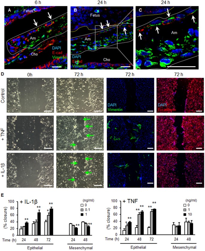
Epithelial-mesenchymal transition (EMT) in mouse pPROM and human amnion epithelial cells. (A–C) Immunostaining for vimentin (green), E-cadherin (red) and DAPI (blue) at ruptured site of amnion past a xx G needle (ø 0.91 mm). Ruptured amnion after 6 h (A, 26 G) and 24 h (B and C, 20 G). (C) is a magnified image of (B). Note that vimentin-positive cells were observed in the epithelial layer of amnion (arrow). Bars, twenty µm. (D and Due east) Wound healing scratch assay of primary human being amnion epithelial cells. Cells were treated with vehicle command, IL-1β (1 ng/ml) or TNF (10 ng/ml) afterward scratching. Phase contrast or immunostaining images with vimentin (dark-green), E-cadherin (red), or DAPI (blue) were obtained at 0 or 72 h. Green arrow indicates mesenchymal-similar shape cells in wounded area of epithelial cells. Bars, 200 µm. (E) Percent closure of scratched human amnion epithelial and mesenchymal cells treated with unlike doses of IL-1β (upper console) or TNF (lower panel). Error confined represent SD. n = 3 in each group. *P < 0.05, **P < 0.01, compared to control (0 ng/ml) at each time point.
Proinflammatory cytokines have potential to induce EMT in healing of skinxvi. Thus, nosotros tested whether IL-1β or TNF induce EMT in principal man amnion epithelial cells. In wound healing scratch assays of amnion epithelial cells, migration was increased dose-dependently by IL-1β or TNF (Fig. 8D,E). In contrast, IL-1β or TNF did not stimulate migration of mesenchymal cells (Fig. 8E). Closure of scratch is considered to be stimulated past migration, not proliferation, of epithelial cells, considering IL-1β or TNF did non change the number of viable cells in proliferation analysis of amnion epithelial cells (information non shown). In addition, IL-6 did not stimulate migration of either epithelial or mesenchymal cells of amnion (data not shown). In the scratched surface area, IL-1β or TNF changed the morphology of amnion epithelial cells into spindle-shaped cells (Fig. 8D, green arrows in second cavalcade). Immunofluorescence revealed that these spindle-shaped cells were vimentin-positive (Fig. 8D, 3rd cavalcade), suggesting EMT, and vimentin-positive cells were also scattered in the not-wounded area (Fig. 8D, third column). Vimentin-positive cells were not observed in control cells without injury. IL-1β and TNF also changed morphology of epithelial cells to spindle-shaped (Fig. 8D, second column). Almost of these cells maintained an epithelial phenotype (i.e., Due east-cadherin positive and vimentin-negative) (Fig. 8D, 3rd and fourth columns) despite the alter in epithelial cell shape. Extension of culture for 5 days resulted in appearance of vimentin-positive cells in the scratched area of primary human being amnion epithelial cells even without cytokine treatment (Fig. S6C). Gene expression demonstrated that vimentin mRNA was significantly increased in scratched cells compared to command cells without injury, whereas East-cadherin mRNA was non inverse (Fig. S6D). Moreover, culture with 10% FBS for 8 days dramatically increased EMT of amnion epithelial cells (Fig. S6A,B). Since FBS includes various cytokines and growth factors, we propose that amnion epithelial cells readily undergo EMT afterward stimulation with these factors. The rate of epithelial cell migration is slower than that of mesenchymal cells (Fig. S5E). EMT would thereby increment the migration rate of epithelial cells thereby accelerating closure of the ruptured amnion. Some of the massive mesenchymal cells aggregating in the healing center in vivo may be derived from cytokine-stimulated amnion epithelial cells through EMT.
Discussion
Here, we investigated the mechanisms of healing of sterile preterm premature rupture of the fetal membranes. Using a preclinical model of pPROM in mice and primary man amnion cells in culture, we demonstrated that (i) sterile pocket-sized rupture of amnion at embryonic 24-hour interval 15 healed spontaneously,(ii) healing of sterile large rupture of amnion was incomplete within 72 h, (iii) Arg1-positive amniotic fluid fetal macrophages were recruited to the site of injury and incorporated into the amnion at after fourth dimension points, and (4) proinflammatory cytokines, IL-1β and TNF, secreted from macrophages recruited at early time points, stimulated EMT of amnion epithelial cells.
Healing of fetal membranes
In our sterile puncture model, we plant that modest ruptures of fetal membranes in mid-gestation (ø 0.47 mm) heal within 3 days. Perchance, this is not surprising in that pocket-size puncture of fetal membranes by amniocentesis in humans heals spontaneously17. Nonetheless, healing of 0.47 mm punctures in mice illustrates that (i) the amnion exhibits hitting regenerative potential18, and (2) embryonic wound healing is rapid and consummate compared with developed tissues7,19. In addition, the surface of migrating amnion mesenchymal cells was covered past a monolayer of epithelial cells. These morphological features were similar to the classical report of sterile rupture of rat fetal membranes in a thickened edge of amnion appeared as a bulge with a pointed tip at 24 h and fibrous spindle-shaped mesenchymal cells were covered past a single layer of flattened epithelial cells20. Still, healing rates of large rupture size (ø 0.91 mm) were simply xl%. This is compatible with clinical findings in which comparably big rupture sizes caused by fetoscopy exercise not heal21. Although choriodecidua does not contribute to the load-begetting chapters of the fetal membranes and not the focus of this investigation, healing of choriodecidua was tedious and incomplete compared with amnion. We speculate that decidual tissue of maternal origin exhibits wearisome rates of healing relative to fetal cells of the amnion. Farther, macrophages readily observed later injury in the amnion were rare subsequently injury of the decidua, suggesting that choriodecidua is less assisted by innate immunity of wound-healing.
Interestingly, amniotic fluid book did non recovered despite closure of the amnion. During normal gestation in the mouse, amniotic fluid decreases significantly during late gestation (from ~120 to 55 µl, Fig. 3C) suggesting that failure to increase amniotic fluid after rupture of the membranes on d15 may merely represent the normal negative balance of fluid accumulation at this time in gestation. This result is also supported by the previous study that amniotic fluid volume decreased significantly in mice from ED 15 to ED 18, different human22, indicating that absorption of amniotic fluid overcomes the synthesis toward term. In addition, although we histologically confirmed closure of amnion, closure of choriodecidua was non consummate at 72 h (Fig. S2). It is possible that lack of full fetal membrane repair resulted in continuous leakage of amniotic fluid. One of the risks of prolonged rupture of the membranes is lack of fetal lung maturity. Examination of fetal lung 3 d later rupture, however, did not reveal negative effects of the modest oligohydramnios during this time in gestation.
Diverse forms of biological materials have been used to repair preterm premature ruptured membranes including fibrin gum, platelet rich plasma, cryoprecipitates, fibrinogen, thrombin, polytetrafluoroethylene material, matrigel, and gelatin sponges23. However the results of these trials were not necessarily satisfactory or remain at the preclinical level. We believe that our mouse pPROM model will be useful for investigating bones mechanisms and treatment of pPROM.
Macrophage infiltration
The presence of macrophages early on later on injury of the amnion was interesting since resident macrophages were not observed in intact amnion and the avascular nature of the amnion would preclude macrophages derived from circulating monocytes. Iii potential sources of amnion macrophages were considered: ane) decidua, 2) amniotic fluid, or three) differentiation of resident amnion stem cells. Advent of macrophages at the site of injury as early equally 2 h of rupture speaks against the possibility of monocyte differentiation which requires several days24. Besides, differentiation of resident stalk cells is unlikely in this time frame. Recruitment of decidual macrophages into amnion was considered considering decidua is enriched in resident macrophages13 However, macrophages were non observed at the wounded choriodecidua. Further, wound-induced accumulation of amnion macrophages were attached to the fetal side of amnion (epithelium) where adhesion molecules P-selectin and VCAM1 were highly expressed but not the adjacent mesenchymal cell layer. Further, these cells were Y positive in the case of a male fetus. Collectively, these results indicate that amniotic fluid is the origin of amnion-healing macrophages. The concept of "migrating amniotic fluid macrophage" is besides reported in other situations during pregnancy25,26. Physiologically, it is known that the number of amniotic fluid macrophages increases toward term in mouse27. In our data, consistent with the previous study, the number of macrophages in amniotic fluid increased half dozen h (ED 15) to 72 h (ED18) (Fig. 7J). These increased amniotic fluid macrophages are considered to infiltrate myometrium and activate myocytes to initiate parturition25,26.
In addition to macrophages, Arg1 was also expressed in amnion epithelial cells. This phenomenon in which Arg1 is highly expressed in inflamed epithelial cells has been reported in other tissues (e.one thousand., lung epithelial cells of patients with asthma28 and skin epidermis of patients with psoriasis29). Since inhibition of Arg1 activity delays cutaneous healing30, we pose that arginase-i is non only a mark of M2 macrophages but also plays a function in healing of the amnion.
In the process of wound healing, macrophages phagocytose organisms and matrix debris, and release various growth factors and cytokines. In this study, we observed secretion of IL-1β and TNF from macrophages which stimulated EMT of amnion epithelial cells. EMT is observed during embryogenesis, organ evolution, wound healing, tissue regeneration, and cancer and its metastasis31. Jail cell-prison cell contacts are of import in the maintenance of an epithelial phenotype. Disruption of cell-cell contacts initiates EMT via cadherin switching32, which resulted in generation of mesenchymal-shaped cells in our mouse model and primary amnion cells. Thus, although macrophages may serve a positive role in healing acutely, excessive or chronic inflammation delays wound healing33. In our model of sterile pPROM, increased expression of the anti-inflammatory cytokine IL-10 accompanied amplified expression of inflammatory cytokines. IL-10 inhibits production of proinflammatory cytokines (e.g., IL-1α, IL-1β, TNF, and IL-half-dozen) from activated macrophage34. Further, treatment of cutaneous wounds with IL-10 neutralizing antibodies results in overexpression of chemokines and cytokines and exaggerated product of neutrophil chemoattractants35. In this ruptured membrane model, secretion of IL-1β and TNF was well-localized and express, suggesting that anti-inflammatory mechanisms (eastward.thousand., IL-10) tightly regulate the inflammatory response to optimize wound healing of amnion. It should also be emphasized that these unique mechanisms become crucial in a tissue that cannot depend on growth factors and other stimulants from the vasculature.
Overall mechanisms of membrane healing: pocket-sized vs large injury
Our mouse models of pPROM betoken that small-scale ruptures heal but the majority of large ruptures practice not. Comparisons betwixt the two models reveal potential mechanisms for these differences. First, cytokine and chemokine responses are transient subsequently modest rupture, but prolonged subsequently larger injury. 2nd, MMP9 gene expression is upregulated dramatically in the later stages of wound healing in the large rupture but not pocket-sized. Third, residual amniotic fluid is present afterwards minor, simply not big, rupture. This amniotic fluid, although decreased relative to intact membranes, may serve as a reservoir for macrophages and other healing factors or their binding proteins. Regardless, the major problem in healing after large rupture seems to be the lack of mesenchymal cell migration and matrix deposition. In the case of pocket-size ruptures, EMT may be sufficient to span matrix support and strength of the membrane. Afterwards large ruptures, nonetheless, it is likely to be insufficient to scaffold the large mesenchymal defect, peculiarly in the presence of protease activation.
Summary
In conclusion, we analyzed in detail spontaneous healing of the fetal amnion using preclinical models of small and big premature rupture of the membranes. We conclude that amnion has high regenerative potential through unique mechanisms including (i) recruitment of fetal macrophages from the amniotic fluid, and (two) IL-1β- and TNF-induced EMT and migration of amnion epithelial cells. Healing of large ruptures is compromised probable due to defects in mesenchymal cell migration and matrix deposition. We suggest, therefore, that the model is an interesting first pace in understanding the role of "sterile inflammation" and healing of the amnion.
Materials and Methods
Preclinical model of sterile ruptured membrane
All animals were handled and euthanized in accordance with the standards of humane beast care described by the National Institutes of Health Guide for the Care and Use of Laboratory Animals, using protocols approved by the Institutional Animal Care and Use Commission (IACUC) of the University of Texas Southwestern Medical Center at Dallas. On xv days postal service coitum (dpc), C57/Bl6 significant mice were anesthetized with isoflurane. A ventral incision was made and uterus and fetal membranes punctured with sterile needles (26 or 20 approximate). In each uterus, half of gestational sacs were punctured (rupture group) and half were not (intact command). In the case of 20 G injury, 4 punctures were conducted per sac whereas eight punctures per sac were conducted with 26 Yard needles. Rupture of membrane was confirmed past leakage of amniotic fluid. To mark puncture sites, needles were soaked in ten% Republic of india Ink before puncture resulting in black staining of the puncture site with contact of the outer surface of the needle. After each procedure, the maternal abdomen was closed with v-0 Polysorb. At the time of sampling, diameter of rupture was measured by a digital caliper under microscopy. Closure was defined equally an invisible hole nether microscopy and the membrane continuous at the rupture site. Fetal membranes were nerveless after washing with PBS, quick frozen in liquid nitrogen, and stored at −80 °C. All procedures adhered to the NIH Guide for the Care and Use of Laboratory.
Scanning Electron Microscope (SEM)
Fetus, fetal membranes, and placenta were fixed with 2.5% (v/five) glutaraldehyde in 0.1 Yard sodium cacodylate buffer overnight at 4 °C. After 3 rinses in 0.one Yard sodium cacodylate buffer, they were post-fixed with 2% osmium tetroxide in the same buffer for 2 h. Samples were rinsed with water and dehydrated with increasing concentration of ethanol, followed by disquisitional point drying (Tousimis Samdri-795). Dried fetal membrane was cut around the placenta, and fetus and placenta were removed. Fetal membranes were mounted on SEM stubs like a "shell" and fixed with the inside of the fetal membrane shell (amnion side) upward. Membranes were sputter coated with gilded/palladium (Cressington 108 machine sputter coater) and images acquired on a Field-Emission Scanning Electron Microscope (Zeiss Sigma) at iii.0 kV accelerating voltage.
Quantitative real-time PCR
Not-injured tissue around the rupture site would dilute the changes of mRNA level, and then we made multiple punctures per gestation sac to increase the ruptured area. In addition, we cut off non-ruptured site of fetal membrane as far as possible at sampling. Quantitative RT-PCR was used to make up one's mind the relative levels of gene expression. Primer sequences for amplifications are shown in Supplementary Table 1. SYBR greenish to detect amplification and gene expression was normalized to that of indicated housekeeping factor.
Isolation and culture of amnion epithelial and mesenchymal cells
Separation and isolation of homo amnion epithelial and mesenchymal cells were performed as previously described36. The protocol for obtaining placentae and fetal membranes was approved by the Institutional Review Board of the University of Texas Southwestern Medical Center. Briefly, amnion tissue was separated by blunt autopsy and then minced. Cells were dispersed by enzymatic digestion with trypsin (Trypsin 1:250, #27250-018, Gibco), and then collagenase (Collagenase B, #11088831001, Roche) and DNase I (10104159001, Roche). Isolated amnion cells were suspended in DMEM/F12 (D8437, Sigma) with x% fetal bovine serum and 1% antibiotic-antimycotic solution (Anti Anti 100X, #15240-062, Gibco). Cells were plated in plastic culture dishes, maintained at 37 °C in a humidified atmosphere of 5% CO2 in air, and allowed to replicate in monolayer to confluence.
Immunofluorescent staining
Mouse uterus was stock-still in 4% paraformaldehyde overnight and kept in fifty% ethanol until paraffin embedding. To ensure that sites of injury were localized, sections were targeted at the black stain left by India Ink needles. Second, series sections were taken every 500 µm throughout the injury, and serial sections of nonruptured sacs were likewise obtained to ensure that the defects were not due to training artefacts. After sections (5 μm) were deparaffinized, antigen retrieval was performed by incubation with proteinase G (P8107S, New England Biolab, working concentration, 0.half dozen units/ml) for 1 infinitesimal at 37 °C. For antigen retrieval of CD68, E-cadherin, and vimentin, sections were boiled in 10 mM sodium citrate buffer (pH 6.0) past microwave for 20 min, and then cooled for 30 min at room temperature. Afterwards antigen retrieval, sections were preincubated with 10% normal goat serum (50062Z, Life Technologies) with 0.3% Triton 10-100 for 30 min at room temperature. Subsequently, tissue sections were incubated with primary antibody in PBS with 1% BSA and 0.3% Triton X-100 at 4 °C overnight. Main antibodies used and concentration were as follows: F4/80 (clone BM8, eBioscience, #14-4801, one:l), Vimentin (D21H3, Prison cell Signaling, #5741, i:100), E-cadherin (4A2, Cell Signaling, #14472, one:50), VCAM1 (EPR5047, Abcam, #134047, 1:100), P-selectin (CD62P, Abcam, #202983, i:100), NOS2 (iNOS, Abcam, #15323, one:100), Arg1 (Arginase-one, D4E3M, Prison cell Signaling, #93668, 1:100), TNF (Abcam, #6671, ane:100), IL-1β (Abcam, #9722, ane:100), Ly-6G (eBioscience, #14-5931, ane:100), CD68 (KP1, Abcam, #955, 1:100), Ki67 (Abcam #15580, 1:100), HSP70 (Abcam #2787, 1:100). Thereafter, sections were incubated with Alexa Fluor 488, 549, or 594–conjugated secondary antibodies (Ig, heavy and lite chains, Invitrogen, 1:500) in 10% normal caprine animal serum for 1 h at room temperature. After that, slides were mounted with Prolong Gold Antifade Reagent with DAPI (P36935, Molecular Probes). Images were taken past Leica SP5 confocal microscopy. ImageJ software was used to generate private images.
Fluorescence in situ hybridization of Y chromosome
Images of sections immunostained with F4/80 and DAPI (antigen retrieval, proteinase G at 37 °C for 1 minute) were obtained earlier hybridization using Leica SP5 confocal microscopy. Thereafter, the encompass drinking glass was removed by shaking in warmed PBS (37 °C, 30 min)., sections were washed by 2X saline-sodium citrate buffer (SSC), and so pretreated with 10 mM sodium citrate (pH vi.0)/2 mM EDTA (pH 8.0) at 80 °C for 45 min. Later on washing with 2X SSC, sections were dehydrated with seventy% ethanol (1 minute) so 100% ethanol (i minute). Dry sections and murine Y chromosome probe (Cytocell, AMP0YR) were pre-warmed at 37 °C for 5 min. 10 µl of FISH probe was practical for 22 mm × 22 mm area, and covered with cover drinking glass and sealed with safety cement. After glue was dry out, sections were denatured on hot-plate at 75 °C for 5 min. Sections were then incubated in humidified box at 37 °C overnight. After removing comprehend glass, sections were done with 2X SSC (room temperature, 5 min), 2X SSC/0.iii% NP-40 (75 °C, 5 min), so 2X SSC (room temperature, i min), and mounted with Prolong Gold Antifade Reagent with DAPI. Images were obtained with Leica SP5 confocal microscopy. The fields of immunostaining and FISH images were matched according to the location of nuclear distribution to be as close to each other as possible37. ImageJ software was used for image processing.
Wound healing scratch assay
Primary human amnion epithelial or mesenchymal cells were harvested in 12-well plate. At confluency, the center of each well was scratched with a 10 µl tip (epithelial cells) or a 200 µl tip (mesenchymal cells). After washing with PBS, IL-1β (#201-LB, R&D Systems), or TNF (#210-TA, R&D Systems) was added in serum-free medium. The width of wound was measured microscopically with 3 points in each field. 3 different fields were selected in each well, and width was averaged. Pct closure was calculated by dividing the average width of the indicated time by the width of 0 h.
Immunocytochemistry
Primary human being amnion epithelial or mesenchymal cells were grown in 8-well sleeping accommodation slide. Cells fixed in four% paraformaldehyde for ten min. Subsequently washing with PBS, slides were incubated with 10% normal goat serum for xxx min at room temperature. Thereafter, slides were incubated with primary antibodies overnight at 4 °C. Primary antibodies used and concentration was as follows: Vimentin (D21H3, Cell Signaling, #5741, 1:100), E-cadherin (4A2, Cell Signaling, #14472, 1:fifty), and CD68 (KP1, Abcam, #955, 1:100). Later on incubation with secondary antibiotic (Alexa Fluor 488, 549, or 594, Invitrogen, ane:500 dilution), slides were mounted with Prolong Golden DAPI. Images were taken past Leica SP5 confocal microscopy, and generated by ImageJ software.
Isolation of mouse amniotic fluid macrophage
Amniotic fluid macrophages were isolated from mice as described previously26. Briefly, amniotic fluid from private sacs was aspirated with a xx-gauge needle with i.0-ml syringe containing 100 µl of RPMI medium 1640. Nerveless amniotic fluid of each gestational sac was added to the 8-well bedroom slide, and incubated at 37 °C for 1 h in incubator. After confirming cell attachment to the slide, cells were fixed with 4% paraformaldehyde for 10 min, and then processed for immunocytochemistry. For quantification of macrophages, five fields were randomly selected using 20× objective and the average number of CD68-positive cells per field was divided past the amniotic fluid volume (µl).
Statistical Analysis
Values were expressed every bit means ± SD in vitro experiments or ways ± SEM in mice experiments. Data were analyzed by unpaired Pupil'south t- test unless indicated otherwise. For comparison of diameter of rupture of amnion and choriodecidua, and number of CD68 positive cells in amniotic fluid, ANOVA was conducted. For complete closure of amnion, chi-square exam was utilized. P-values less than 0.05 were regarded as statistically pregnant.
References
-
Parry, S. & Strauss, J. F. third Premature rupture of the fetal membranes. N Engl J Med 338, 663–670, https://doi.org/10.1056/NEJM199803053381006 (1998).
-
Goldenberg, R. 50., Culhane, J. F., Iams, J. D. & Romero, R. Epidemiology and causes of preterm nativity. Lancet 371, 75–84, https://doi.org/10.1016/S0140-6736(08)60074-iv (2008).
-
Romero, R. et al. Intraamniotic infection and the onset of labor in preterm premature rupture of the membranes. Am J Obstet Gynecol 159, 661–666 (1988).
-
Gomez, R. et al. The fetal inflammatory response syndrome. Am J Obstet Gynecol 179, 194–202 (1998).
-
Johnson, J. West., Egerman, R. S. & Moorhead, J. Cases with ruptured membranes that "reseal". Am J Obstet Gynecol 163, 1024–1030; discussion 1030–1022 (1990).
-
Martin, P. Wound healing–aiming for perfect skin regeneration. Science 276, 75–81 (1997).
-
Sonnemann, K. J. & Bement, West. K. Wound repair: toward understanding and integration of unmarried-cell and multicellular wound responses. Annu Rev Cell Dev Biol 27, 237–263, https://doi.org/10.1146/annurev-cellbio-092910-154251 (2011).
-
Gordon, S. Alternative activation of macrophages. Nat Rev Immunol 3, 23–35, https://doi.org/ten.1038/nri978 (2003).
-
Gordon, Southward. & Martinez, F. O. Alternative activation of macrophages: mechanism and functions. Immunity 32, 593–604, https://doi.org/10.1016/j.immuni.2010.05.007 (2010).
-
Murray, P. J. & Wynn, T. A. Protective and pathogenic functions of macrophage subsets. Nat Rev Immunol 11, 723–737, https://doi.org/x.1038/nri3073 (2011).
-
Underwood, M. A., Gilbert, W. G. & Sherman, M. P. Amniotic fluid: not just fetal urine anymore. J Perinatol 25, 341–348, https://doi.org/10.1038/sj.jp.7211290 (2005).
-
Sutherland, G. R., Bauld, R. & Bain, A. D. Observations on human being amniotic fluid cell strains in serial culture. J Med Genet 11, 190–195 (1974).
-
Sutton, L., Mason, D. Y. & Redman, C. W. HLA-DR positive cells in the human placenta. Immunology 49, 103–112 (1983).
-
Shaw, T. J. & Martin, P. Wound repair at a glance. J Cell Sci 122, 3209–3213, https://doi.org/x.1242/jcs.031187 (2009).
-
Imhof, B. A. & Aurrand-Lions, M. Adhesion mechanisms regulating the migration of monocytes. Nat Rev Immunol iv, 432–444, https://doi.org/x.1038/nri1375 (2004).
-
Yan, C. et al. Epithelial to mesenchymal transition in human peel wound healing is induced past tumor necrosis factor-alpha through bone morphogenic protein-2. Am J Pathol 176, 2247–2258, https://doi.org/ten.2353/ajpath.2010.090048 (2010).
-
Borgida, A. F., Mills, A. A., Feldman, D. 1000., Rodis, J. F. & Egan, J. F. Issue of pregnancies complicated by ruptured membranes after genetic amniocentesis. Am J Obstet Gynecol 183, 937–939, https://doi.org/10.1067/mob.2000.108872 (2000).
-
Maxson, S., Lopez, E. A., Yoo, D., Danilkovitch-Miagkova, A. & Leroux, K. A. Concise review: role of mesenchymal stem cells in wound repair. Stalk Cells Transl Med one, 142–149, https://doi.org/10.5966/sctm.2011-0018 (2012).
-
Redd, Yard. J., Cooper, Fifty., Wood, Due west., Stramer, B. & Martin, P. Wound healing and inflammation: embryos reveal the manner to perfect repair. Philos Trans R Soc Lond B Biol Sci 359, 777–784, https://doi.org/10.1098/rstb.2004.1466 (2004).
-
Sopher, D. The response of rat fetal membranes to injury. Ann R Coll Surg Engl 51, 240–249 (1972).
-
Gratacos, Eastward. et al. A histological study of fetoscopic membrane defects to certificate membrane healing. Placenta 27, 452–456, https://doi.org/x.1016/j.placenta.2005.03.008 (2006).
-
Cheung, C. Y. & Brace, R. A. Amniotic fluid volume and limerick in mouse pregnancy. Journal of the Society for Gynecologic Investigation 12, 558–562, https://doi.org/x.1016/j.jsgi.2005.08.008 (2005).
-
Devlieger, R., Millar, 50. G., Bryant-Greenwood, G., Lewi, L. & Deprest, J. A. Fetal membrane healing after spontaneous and iatrogenic membrane rupture: a review of current evidence. Am J Obstet Gynecol 195, 1512–1520, https://doi.org/x.1016/j.ajog.2006.01.074 (2006).
-
Geissmann, F. et al. Development of monocytes, macrophages, and dendritic cells. Science 327, 656–661, https://doi.org/ten.1126/scientific discipline.1178331 (2010).
-
Thomson, A. J. et al. Leukocytes infiltrate the myometrium during human parturition: farther evidence that labour is an inflammatory process. Hum Reprod 14, 229–236 (1999).
-
Condon, J. C., Jeyasuria, P., Faust, J. M. & Mendelson, C. R. Surfactant protein secreted by the maturing mouse fetal lung acts as a hormone that signals the initiation of parturition. Proc Natl Acad Sci Us 101, 4978–4983, https://doi.org/ten.1073/pnas.0401124101 (2004).
-
Kobayashi, Grand., Umezawa, One thousand. & Yasui, M. Apoptosis in mouse amniotic epithelium is induced by activated macrophages through the TNF receptor type 1/TNF pathway. Biol Reprod 84, 248–254, https://doi.org/ten.1095/biolreprod.110.087577 (2011).
-
North, M. L., Khanna, N., Marsden, P. A., Grasemann, H. & Scott, J. A. Functionally important role for arginase 1 in the airway hyperresponsiveness of asthma. Am J Physiol Lung Cell Mol Physiol 296, L911–920, https://doi.org/10.1152/ajplung.00025.2009 (2009).
-
Bruch-Gerharz, D. et al. Arginase 1 overexpression in psoriasis: limitation of inducible nitric oxide synthase activeness equally a molecular mechanism for keratinocyte hyperproliferation. Am J Pathol 162, 203–211, https://doi.org/10.1016/S0002-9440(10)63811-4 (2003).
-
Campbell, 50., Saville, C. R., Murray, P. J., Cruickshank, S. 1000. & Hardman, G. J. Local arginase 1 activity is required for cutaneous wound healing. J Invest Dermatol 133, 2461–2470, https://doi.org/x.1038/jid.2013.164 (2013).
-
Kalluri, R. & Weinberg, R. A. The nuts of epithelial-mesenchymal transition. J Clin Invest 119, 1420–1428, https://doi.org/10.1172/JCI39104 (2009).
-
Wheelock, One thousand. J., Shintani, Y., Maeda, One thousand., Fukumoto, Y. & Johnson, K. R. Cadherin switching. J Cell Sci 121, 727–735, https://doi.org/10.1242/jcs.000455 (2008).
-
Eming, South. A., Martin, P. & Tomic-Canic, M. Wound repair and regeneration: mechanisms, signaling, and translation. Sci Transl Med six, 265sr266, https://doi.org/10.1126/scitranslmed.3009337 (2014).
-
Moore, 1000. W., de Westward Malefyt, R., Coffman, R. L. & O'Garra, A. Interleukin-10 and the interleukin-10 receptor. Annu Rev Immunol 19, 683–765, https://doi.org/ten.1146/annurev.immunol.xix.1.683 (2001).
-
Sato, Y., Ohshima, T. & Kondo, T. Regulatory role of endogenous interleukin-10 in cutaneous inflammatory response of murine wound healing. Biochem Biophys Res Commun 265, 194–199, https://doi.org/x.1006/bbrc.1999.1455 (1999).
-
Casey, M. 50. & MacDonald, P. C. Interstitial collagen synthesis and processing in human amnion: a property of the mesenchymal cells. Biol Reprod 55, 1253–1260 (1996).
-
Miyata, Eastward. et al. Hematopoietic origin of hepatic stellate cells in the developed liver. Blood 111, 2427–2435, https://doi.org/ten.1182/claret-2007-07-101261 (2008).
Acknowledgements
Nosotros thank Mr. Jesus' Acevedo and Ms. Haolin Shi for pregnant mice training, the physicians and staff of Parkland Memorial Infirmary, and Ms. Sylvia Wright for valuable aid in tissue procurement. We also thank the shared resource cores in Microscopic Imaging, Molecular Pathology (Mr. John Shelton), Electron Microscopy (Dr. Anza Darehshouri and Ms. Phoebe Doss) at UTSWMC for technical support. This work was supported by March of Dimes Foundation Premature Research Initiation grants #21-FY13–35 and #21-FY15-138. The Tissue Conquering Core supported by NIH P01HD087150 is gratefully best-selling.
Author data
Affiliations
Contributions
H.M. conducted all experiments. A.H.G. assisted H.Thou. in experiments related cell civilization experiments. Y.A. assisted in immunofluorescent imaging of fetal membranes. H.M. and R.A.Westward. wrote the principal manuscript text and prepared all figures. All authors reviewed the manuscript.
Respective author
Ideals declarations
Competing Interests
The authors declare that they accept no competing interests.
Additional information
Publisher'southward annotation: Springer Nature remains neutral with regard to jurisdictional claims in published maps and institutional affiliations.
Electronic supplementary material
Rights and permissions
Open Access This commodity is licensed nether a Creative Commons Attribution iv.0 International License, which permits use, sharing, adaptation, distribution and reproduction in any medium or format, as long every bit yous requite appropriate credit to the original author(s) and the source, provide a link to the Creative Commons license, and indicate if changes were fabricated. The images or other tertiary party fabric in this article are included in the article's Creative Commons license, unless indicated otherwise in a credit line to the material. If material is not included in the article's Creative Eatables license and your intended use is non permitted past statutory regulation or exceeds the permitted use, you lot will need to obtain permission directly from the copyright holder. To view a copy of this license, visit http://creativecommons.org/licenses/by/4.0/.
Reprints and Permissions
About this article
Cite this article
Mogami, H., Hari Kishore, A., Akgul, Y. et al. Healing of Preterm Ruptured Fetal Membranes. Sci Rep seven, 13139 (2017). https://doi.org/ten.1038/s41598-017-13296-i
-
Received:
-
Accepted:
-
Published:
-
DOI : https://doi.org/10.1038/s41598-017-13296-ane
Further reading
Comments
By submitting a comment yous agree to abide by our Terms and Customs Guidelines. If you find something abusive or that does not comply with our terms or guidelines delight flag it as inappropriate.
Source: https://www.nature.com/articles/s41598-017-13296-1
Posted by: duvallfaight.blogspot.com


0 Response to "Is It Possible For The Body To Repair A Small Leak In Amniotic Fluid"
Post a Comment